

If you could look at the back of a healthy human eye with the right magnifying instrument, you would see something remarkable at the center of the retina: a subtle but distinct golden-yellow spot. That spot is the macula, named from the Latin for “spot,” and its yellowish tint comes from a layer of pigment that sits within its inner layers, absorbing certain wavelengths of light and giving the region its characteristic color. That pigment, the macular pigment, is one of the most functionally important structures in the entire visual system, and it’s built almost entirely from what you eat.
Most people have never heard of the macular pigment. Fewer still know that its density, directly measurable and highly variable between individuals, is one of the best-supported predictors of both current visual performance and long-term retinal health that nutritional science has identified. Getting to grips with what the macular pigment is and how to support it is one of the most practical things you can learn about caring for your eyes.
Contents
What the Macular Pigment Is Made Of
The macular pigment is composed of three carotenoid compounds: lutein, zeaxanthin, and meso-zeaxanthin. All three are members of the xanthophyll branch of the carotenoid family, and all three are characterized by the yellow-orange color that gives the pigment its appearance.
Of these three, only lutein and zeaxanthin are provided directly by diet. Meso-zeaxanthin is not found in meaningful amounts in any common food. Instead, the retina produces it locally through metabolic conversion from lutein, using the lutein that arrives from dietary absorption as raw material. This means that while meso-zeaxanthin is a significant component of the macular pigment, particularly at the foveal center, the supply chain for it begins in the kitchen or the supplement cabinet rather than in the macula itself.
Where Each Compound Concentrates
The three macular carotenoids don’t distribute uniformly across the macula. Meso-zeaxanthin and zeaxanthin concentrate most densely at the foveal center, the tiny pit of maximum cone photoreceptor density responsible for the sharpest central vision. Lutein’s distribution is broader, extending throughout the peripheral macula in a ring around the foveal core. This spatial arrangement creates a pigment whose density is greatest precisely where the retina is most metabolically active and most vulnerable to photo-oxidative damage.
The binding proteins that capture and hold these carotenoids in the macular tissue, called GSTP1 (glutathione S-transferase pi 1) for zeaxanthin and meso-zeaxanthin and StAR-D3 for lutein, are expressed specifically by the Muller cells and the retinal pigment epithelium cells of the macula. Their selective expression in this region is what causes the extraordinary accumulation of carotenoids here compared to any other tissue in the body.
What the Macular Pigment Does
The macular pigment performs its protective functions through two distinct but complementary mechanisms that together constitute the retina’s primary nutritional defense against light-induced damage.
Optical Blue Light Filtration
The absorption spectrum of the macular pigment carotenoids peaks in the blue-violet range of visible light, roughly 430 to 490 nanometers. This isn’t coincidental. Blue light carries more energy per photon than longer visible wavelengths and generates reactive oxygen species more efficiently when absorbed by retinal tissue. It also scatters more within the eye, creating optical noise called chromatic aberration that slightly degrades image quality.
By absorbing this high-energy, high-scatter portion of the incoming light before it reaches the photoreceptors, the macular pigment performs two functions simultaneously: it reduces the oxidative damage potential of the light reaching the photoreceptors, and it improves the optical quality of the retinal image by removing the component most responsible for blurring and contrast reduction. Higher macular pigment density means more filtration, better protection, and measurably better visual performance.
Chemical Antioxidant Activity
Beyond its optical filtering role, the macular pigment also serves as a chemical antioxidant in situ. Lutein and zeaxanthin within the pigment layer can directly quench singlet oxygen and neutralize other reactive species generated in the macular tissue by photon absorption and normal metabolic activity. This chemical defense is the last line of protection for the photoreceptors, catching the reactive molecules that get past the optical filter before they can oxidize cellular structures.
The combination of optical pre-filtration and chemical antioxidant activity makes the macular pigment a more complete protective system than either function alone could provide. It is, in the most literal sense, the eye’s built-in sunscreen, operating continuously, wavelength-selectively, and at the exact location of greatest need.
Macular Pigment Ocular Density: The Measurable Metric
Macular pigment density is not fixed. It varies between individuals by as much as an order of magnitude, meaning some people have ten times the macular pigment of others, and it changes within an individual in response to dietary intake, age, and lifestyle factors. This variability is measured as Macular Pigment Ocular Density (MPOD), and it’s one of the more clinically informative measures available in eye health.
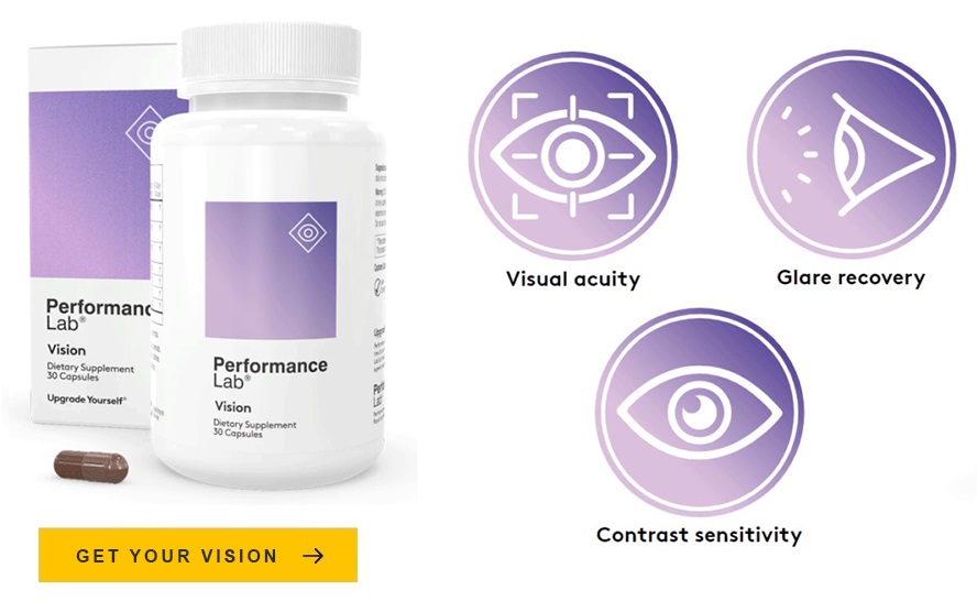
Higher MPOD is consistently associated with better visual acuity, better contrast sensitivity, faster glare recovery, and significantly reduced risk of age-related macular degeneration. Lower MPOD, particularly below 0.3 optical density units, is considered a risk factor for AMD and is associated with measurably poorer performance on visual function tests. The AREDS2 clinical trial established that lutein and zeaxanthin supplementation producing MPOD increases reduced the risk of advanced AMD progression by approximately 26% in high-risk individuals.
What Depletes Macular Pigment
Several factors work against the macular pigment’s density, and understanding them explains why so many adults have lower MPOD than would be optimal.
Dietary insufficiency is the primary driver. Lutein and zeaxanthin cannot be synthesized by the body, and without consistent dietary intake, the pigment cannot be replenished as it naturally turns over. Most Western diets, heavy on processed and refined foods and light on dark leafy greens and eggs, provide far less lutein and zeaxanthin than the amounts that support meaningful macular pigment maintenance. Estimated average dietary intakes of 1 to 2 milligrams per day contrast with the 10 or more milligrams per day used in studies demonstrating significant MPOD increases.
Smoking dramatically reduces MPOD, both by impeding carotenoid absorption and by dramatically increasing the oxidative burden on retinal tissue, depleting the macular carotenoids faster than they can be replaced. Obesity, through sequestration of fat-soluble carotenoids in adipose tissue, reduces their availability to the retina. Chronic high blue light exposure without adequate nutritional support accelerates pigment depletion. And advancing age reduces the efficiency of carotenoid absorption and transport, making adequate dietary intake progressively more important as people get older.
Building the Pigment Through Nutrition
The constructive news is that macular pigment responds reliably to nutritional intervention. Studies consistently demonstrate that supplementation with lutein and zeaxanthin at meaningful doses, typically 10 milligrams of lutein and 2 milligrams of zeaxanthin per day, produces measurable MPOD increases over weeks to months of consistent intake. The response is not immediate, because the macular pigment is a structural component that builds gradually from circulating carotenoids, but it is reliable and sustained with continued supplementation.
The practical implications are worth stating plainly. Macular pigment is the foundation of the retina’s primary protective system. It’s built from dietary carotenoids that most people don’t consume in adequate amounts. It can be meaningfully increased through consistent nutritional support. And the evidence that higher macular pigment density translates into better vision and lower AMD risk is among the most robustly supported in nutritional ophthalmology. There are few investments in eye health with a clearer evidence base, or with a more direct connection between what you eat and what you see.