

In nutrition science, most compounds get studied in isolation first and in combination later. Researchers identify a promising molecule, test what it does on its own, publish the results, and eventually, if things go well, investigate how it interacts with other nutrients. Lutein and zeaxanthin have followed a slightly different arc. While both have been studied individually, the most compelling and clinically significant research has almost always examined them together, and for good reason. These two carotenoids don’t just happen to coexist in the macula. They work as a genuine partnership, each one covering territory the other doesn’t, in ways that make the combination substantially more valuable than either compound in isolation.
Understanding the lutein-zeaxanthin partnership means understanding both what they share and what makes each of them distinct. Their similarities explain why they accumulate in the same location. Their differences explain why both are necessary.
Contents
What They Have in Common
Lutein and zeaxanthin are both xanthophyll carotenoids, members of the broad pigment family that includes hundreds of related compounds found throughout the plant and animal kingdoms. Both are fat-soluble, meaning they require dietary fat for proper absorption. Both are found in similar food sources, principally dark leafy greens and egg yolks. Both are absorbed from the digestive tract and transported in the blood before being selectively deposited in the macula by specific binding proteins that the eye produces precisely for this purpose.
Most importantly, both absorb blue light. The yellow-orange color of the macular pigment they form together is a visual signature of this property: yellow-orange pigments absorb the blue end of the light spectrum and reflect the warmer wavelengths. When blue light enters the eye, a significant portion of it encounters the macular pigment before reaching the photoreceptors, and a significant portion of that is absorbed and neutralized before it can generate damaging free radicals in the sensitive cells beneath.
Structural Similarity, Functional Distinction
Chemically, lutein and zeaxanthin are almost identical. They share the same molecular formula (C40H56O2) and differ only in the position of a single double bond in their ring structures. This is a modest structural difference, but it’s enough to give them somewhat different optical properties and, more importantly, different affinities for different parts of the macula. That spatial distinction is the key to understanding why the partnership matters.
How They Divide the Macular Territory
The macula is not a uniform structure. It’s a layered region of the central retina with distinct anatomical zones, and the distribution of lutein and zeaxanthin across these zones reflects their complementary roles.
Zeaxanthin’s Domain: The Foveal Center
Zeaxanthin is preferentially concentrated at the fovea, the central pit of the macula where cone photoreceptors are most densely packed and where visual acuity is sharpest. At the very center of the fovea, zeaxanthin can account for the majority of the total macular carotenoid content. This is the region that receives the most focused, concentrated light and therefore bears the highest risk of photo-oxidative damage. Zeaxanthin’s disproportionate presence at this most critical location reflects its particular affinity for the specialized binding proteins expressed by the foveal Muller cells.
The practical implication is direct: zeaxanthin is the primary nutritional guardian of the photoreceptors responsible for your sharpest, most detailed central vision. Adequate zeaxanthin availability is what determines whether the fovea has the protective pigment density it needs at its most vulnerable center.
Lutein’s Domain: The Peripheral Macula
Lutein’s distribution is broader, extending throughout the peripheral macula in a ring around the foveal center. In absolute amounts, the macula actually contains more lutein than zeaxanthin because lutein’s territory is larger, even though zeaxanthin is more concentrated at the foveal tip. Lutein provides blue light filtration and antioxidant protection across the wider macular region, covering the transition zone between the high-acuity fovea and the surrounding retina.
A third compound, meso-zeaxanthin, is also present in the macula, particularly in the foveal region. Meso-zeaxanthin is not found in significant amounts in food; it is produced in the retina from lutein through metabolic conversion. Its presence reinforces the foveal carotenoid concentration, and inadequate lutein intake, by limiting the substrate available for this conversion, can reduce meso-zeaxanthin levels and leave the fovea more vulnerable.
Their Combined Effect on the Macular Pigment
Together, lutein, zeaxanthin, and meso-zeaxanthin form the macular pigment, the yellow filter layer that is the retina’s primary defense against light-induced oxidative damage. The density of this pigment, measured as Macular Pigment Ocular Density (MPOD), determines the eye’s level of blue light protection and correlates with multiple measures of visual performance and long-term retinal health.
MPOD responds to both lutein and zeaxanthin intake, but the two compounds contribute to different spatial regions of the pigment. A supplement strategy that provides only lutein will increase the peripheral macular pigment but may leave foveal zeaxanthin density suboptimal. One that provides only zeaxanthin will reinforce the foveal center but leave the broader macular region with less coverage. The combination, reflecting the natural co-occurrence of these compounds in the macula, provides comprehensive protection across the entire macular region.
The AREDS2 clinical trial, which followed more than 4,000 high-risk participants, found that supplementing with lutein and zeaxanthin together reduced the risk of progression to advanced age-related macular degeneration by approximately 26%. This remains the landmark evidence for the combined protective value of these carotenoids, and it was achieved with the combination, not with either compound alone.
Visual Performance Benefits of the Duo
Beyond long-term macular protection, the lutein-zeaxanthin partnership produces measurable improvements in how the eyes actually perform.
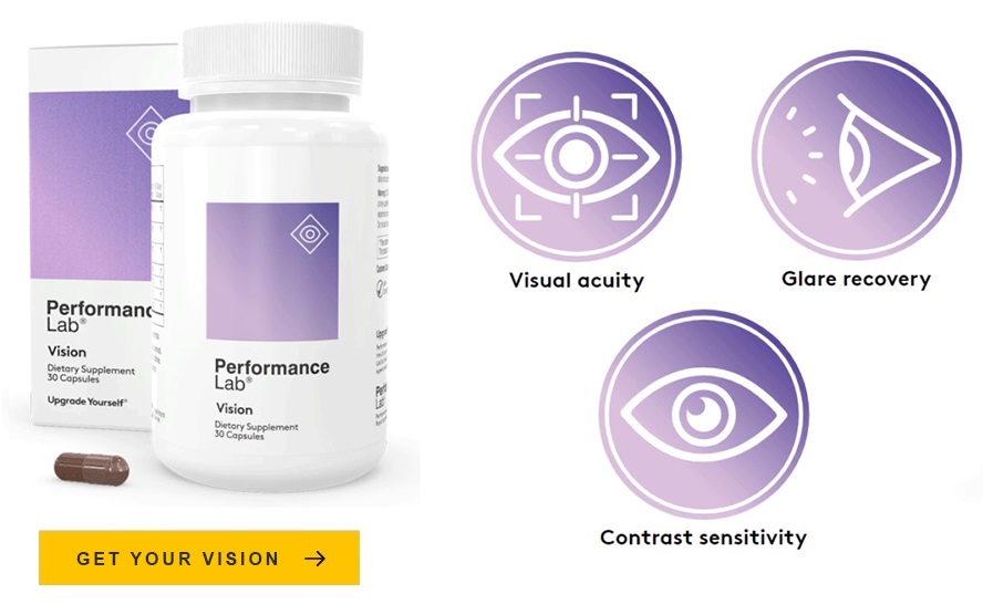
Visual Acuity and Contrast
By filtering blue light scatter, which is the component of visible light most responsible for optical noise and chromatic aberration in the retinal image, the macular pigment improves both visual acuity and contrast sensitivity. Higher MPOD is consistently associated with better performance on both measures. Studies supplementing participants with lutein and zeaxanthin and documenting MPOD increases have also found corresponding improvements in tested acuity and contrast sensitivity scores.
Glare Recovery
The macular pigment’s blue light filtration also accelerates recovery from glare events, the temporary visual disruption caused by intense light sources. Research linking higher MPOD to faster glare recovery reflects the contribution of both carotenoids to the pigment density that makes this function possible. Drivers, athletes, and anyone who moves regularly between bright and dim environments have practical performance reasons to maintain adequate combined intake.
The Dietary and Supplemental Picture
The foods that supply lutein and zeaxanthin in the highest amounts are dark leafy greens (for lutein) and eggs and orange bell peppers (for zeaxanthin). A diet consistently rich in these foods will provide both carotenoids, though the relative proportions will vary depending on which foods dominate. Typical Western diets, heavy on processed foods and light on leafy greens, tend to provide both compounds at levels substantially below those associated with meaningful macular benefits in research.
Supplement formulations for eye health typically provide more lutein than zeaxanthin, in ratios ranging from 5:1 to 10:1, which broadly reflects the natural proportion in which these compounds appear in food sources and in the macula overall. These ratios are not arbitrary; they reflect decades of research on what proportions support the most effective macular pigment building across both the foveal and peripheral macula.
Fat solubility matters for both. Neither lutein nor zeaxanthin will be absorbed efficiently from a supplement or a meal taken without dietary fat. The habit of taking eye health supplements with a meal containing healthy fats is not a minor detail; it’s a prerequisite for getting meaningful amounts of these carotenoids to the retina where they do their work.
A Partnership the Eye Designed
It’s worth pausing to appreciate that the eye didn’t choose lutein and zeaxanthin arbitrarily. Out of the roughly 700 carotenoids found in nature and the 20 or so that circulate in human blood, the macula selectively concentrates these two, and then meso-zeaxanthin derived from them, with specificity that implies profound biological intentionality. The binding proteins that capture them in the macular tissue, the spatial arrangement that places zeaxanthin at the fovea and lutein in the surrounding ring, the ratio in which they accumulate: all of this speaks to a protective architecture that evolution refined over millions of years.
What nutritional science has done is identify the dietary inputs that determine whether that architecture is well-built or compromised. The answer involves making sure both members of the partnership are present in adequate amounts, consistently, over a lifetime. That’s not a complicated strategy. But for the long-term health of the most important sensory organ you have, it may be one of the most valuable ones available.