
Ask most people what’s inside a joint and they’ll say cartilage. Press them a little and they might add tendons and ligaments. Ask them what the difference is between a tendon and a ligament, and the conversation gets interesting. These three tissue types, cartilage, tendons, and ligaments, are the primary structural players in joint health, each performing a distinct and irreplaceable role in allowing movement, absorbing force, and maintaining the stability that makes everyday activity possible without injury. Understanding what each one does, how each one fails, and what each one responds to is fundamental to taking joint health seriously. It’s also less complicated than it sounds once the terminology is out of the way.
Contents
Cartilage: The Cushion and the Bearing Surface
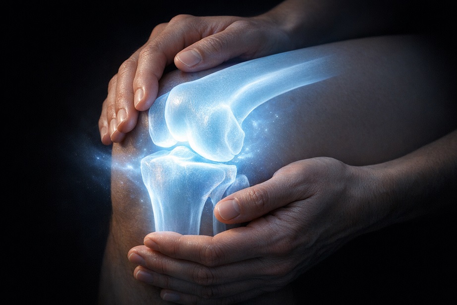
Articular cartilage is the smooth, bluish-white tissue that covers the ends of bones inside synovial joints. It performs two essential functions simultaneously: it reduces friction between opposing bone surfaces to near zero, allowing them to glide across each other with remarkable ease, and it absorbs and redistributes the mechanical forces generated by movement, protecting the underlying bone from the stress concentrations that would otherwise cause damage over time.
Cartilage is composed predominantly of Type II collagen fibers that provide tensile strength and a proteoglycan matrix, primarily aggrecan, that attracts and retains water. This water content, which makes up 65 to 80 percent of healthy cartilage by weight, is what gives the tissue its shock-absorbing compressive properties. When a joint is loaded, water is squeezed from the proteoglycan matrix under pressure, creating hydraulic resistance that distributes the load across the joint surface. When the load is released, the proteoglycans’ strong affinity for water draws it back in, restoring cartilage to its original thickness and readiness for the next loading cycle.
Cartilage’s Critical Vulnerability
What makes cartilage uniquely vulnerable is that it has no blood supply and no lymphatic drainage. Nutrients and oxygen reach it exclusively through diffusion from synovial fluid, driven by the pumping action of joint movement. This means cartilage cannot mobilize the standard vascular repair response that heals other tissues after damage. Its sole maintenance workers are chondrocytes, sparse cells that make up only one to five percent of cartilage volume and become progressively less productive with age. Damage that exceeds chondrocytes’ slow repair capacity tends to accumulate and progress rather than heal. This is the central biological fact that makes cartilage preservation, rather than cartilage regeneration, the realistic goal of joint health strategy.
Ligaments: The Joint’s Structural Constraints
Ligaments are fibrous bands of dense connective tissue that connect bone to bone across joints. They are the structural constraints that define the safe range of motion for each joint, preventing the excessive movement that would stress joint surfaces unevenly or allow bones to dislocate from their articulating positions. The anterior and posterior cruciate ligaments of the knee prevent excessive forward and backward translation of the tibia relative to the femur. The medial and lateral collateral ligaments resist sideways bending stress. Similar arrangements at every major joint in the body reflect the same principle: movement guided and bounded by tensile connective tissue that can resist force without requiring active muscular contraction.
Ligaments are composed predominantly of Type I collagen, arranged in parallel fiber bundles aligned with the primary direction of mechanical loading. This organization gives ligaments their extraordinary tensile strength along the axis of loading, though it also means they are less resistant to forces applied at angles outside that primary direction. Embedded within the collagen matrix are fibroblasts that maintain the tissue, along with small amounts of elastin that contribute the modest elasticity ligaments possess, and proteoglycans and glycosaminoglycans that determine the tissue’s viscoelastic behavior under dynamic loading.

How Ligaments Fail and What They Need
Ligament injuries range from mild stretching with intact fibers (grade 1 sprains) through partial tears (grade 2) to complete rupture (grade 3). Acute sprains typically result from forces that exceed the ligament’s tensile capacity in a sudden, unexpected movement. Chronic ligament problems can also develop from repetitive overload that accumulates microtrauma faster than fibroblasts can repair it, a pattern called ligamentosis that differs from acute sprain both in mechanism and in management approach.
Ligaments age in ways that significantly increase injury vulnerability: collagen production slows, fiber organization becomes less parallel and more random, glycation-based crosslinks accumulate making collagen stiffer and more brittle, and water content declines as glycosaminoglycan concentration falls. Nutrition that supports collagen synthesis, including adequate vitamin C as an essential cofactor, and sulfur from sources like MSM for glycosaminoglycan maintenance, directly addresses these age-related vulnerabilities. The muscle strength surrounding a joint also matters enormously for ligament protection: strong muscles absorb and redirect forces before they reach the passive ligament system, dramatically reducing ligament injury risk under the same external load conditions.
Tendons: The Force Transmitters
Tendons connect muscle to bone, and their role is to transmit the contractile force generated by muscle to the skeletal system that moves in response to it. Every deliberate movement you make involves tendons doing this work, and the forces involved are substantial. The Achilles tendon, for example, routinely experiences loads of several times body weight during running, managed by the extraordinary tensile strength of its densely packed Type I collagen structure.
Like ligaments, tendons are composed primarily of Type I collagen in parallel fiber arrays, maintained by fibroblast-like cells called tenocytes. They have a somewhat better blood supply than ligaments, particularly in their mid-substance, but still significantly less than muscles or bone, which contributes to their relatively slow healing when damaged. Tendons also have a viscoelastic character, meaning they exhibit both elastic (spring-like) and viscous (rate-dependent) behaviors under loading. This viscoelasticity allows tendons to store and release elastic energy during movements like running and jumping, improving the metabolic efficiency of locomotion.
Tendon Injuries and Their Drivers
Tendon injuries span from acute rupture under extreme load to the far more common chronic tendinopathy, a degenerative condition driven by repetitive overloading without adequate recovery that results in disorganized collagen, increased tenocyte cell death, and neovascularization of tissue that is normally relatively avascular. Tendinopathy is perhaps the most common form of overuse injury in active adults and is notoriously slow to resolve because the poor vascular supply of tendon tissue limits the delivery of repair resources.
The nutritional factors that support tendon health overlap significantly with those supporting ligament health. Vitamin C for collagen synthesis, sulfur from MSM for crosslinking and glycosaminoglycan maintenance, and protein adequate for tenocyte collagen production are all relevant. Mechanical loading through progressive exercise is also essential: tendons adapt positively to controlled tensile loading by remodeling their collagen architecture to better resist the demanded forces, and complete rest typically worsens chronic tendinopathy rather than improving it.
A Unified Protection Strategy
What emerges from understanding cartilage, ligaments, and tendons as distinct tissues is that their protection strategies share a coherent core while having tissue-specific emphases. All three depend on collagen and its supporting nutrients. All three are vulnerable to the degrading effects of chronic inflammation and oxidative stress. All three need appropriate mechanical loading to stay healthy and adapt positively to demand. And all three are served by the anti-inflammatory dietary and supplemental strategies that reduce the inflammatory environment accelerating their respective degradation pathways.
The tissue-specific nuances, the need to emphasize cartilage hydration through proteoglycan support, the importance of muscular support for ligaments, the progressive loading protocols specific to tendon rehabilitation, add valuable detail to this unified picture. But the foundational insight is that joint health is not a single-tissue concern. The three primary structural components of joints are interconnected in their function and overlapping in their nutritional and lifestyle needs, which means protecting all three is considerably more achievable than it might initially appear.